
Monoclonal Antibodies (mAbs) analysis Capillary Electrophoresis with Laser Induced Fluorescence Detection
Abstract
Antibody pharmaceuticals is a category of therapeutics that plays an important role in controlling many types of diseases such as cancer, allergy, inflammation, infectious or autoimmune diseases. Monoclonal antibodies have become a fast growing class of biopharmaceutical products.
There is an increased demand in the pharmaceutical industry for tools applicable to large scale production and there is an increasing need for product characterization and control of the manufacturing process. In the biopharmaceutical industry, to support analytical characterization, process development, and quality control of therapeutic antibodies, capillary electrophoresis-sodium dodecyl sulfate (CE-SDS) has been recognized as an important alternative tool of SDS-Page because of the ease of use and the ability to automate.
UV absorbance detection is widely used in CE-SDS. In contrast to standard Liquid Chromatography where proteins are usually detected at 280nm, detector signal in a CE system is not satisfactory due to a very short detection path length (25 to 75µm). Although the absorbance of protein at 210nm is greater than at 280nm, detection in the low UV region is not very sensitive. Alternatively, CE derivatization techniques for Laser Induced Fluorescence detection have been tested to increase detection sensitivity of proteins but the sample preparation is still very restrictive.
In this application note, we present 2 different LIF detections: Fluorescence derivatization and native fluorescence.
Fluorescence Derivatization in CE SDS with LIF
CE derivatization techniques for Laser Induced Fluorescence detection is highly sensitive in protein analysis. But the sample preparation requires purification of labeled mAbs and the derivatizated sample form is unstable.
FITC labeled IgG
Instruments | Agilent HP3D CE Picometrics ZETALIF detector. Laser: 488 nm (excitation) |
Sample | Mouse IgG2a FITC conjugate |
LIF Capillary | Internal Diameter 50µm Total length: 33 cm Effective length: 14 cm |
Method | Injection: sample : 25 seconds at -15kV Analysis condition: -20kV Temperature (cassette and sample): 25°C |
TAMRA labeled IgG
Instruments | Agilent HP3D CE Picometrics ZETALIF detector. Laser: 488 nm (excitation) |
Sample | Tetramethylrhodamine goat anti-mouse IgGMouse IgG |
LIF Capillary | Internal Diameter 50µm Total length: 33 cm Effective length: 14 cm |
Method | Injection: sample : 25 seconds at -15kV Analysis condition: -20kV Temperature (cassette and sample): 25°C |



LOD* obtained for FITC labeled IgG is: 150 pg/mL
- * LOD calculated for a ratio S/N=3
- [1] Anal.Chem. 2006, 78, 6583-6594
LOD* obtained for TAMRA labeled IgG is: 50 pg/mL
When the sample is labeled with TAMRA, preparation and purification of the samples requires many steps [1]: Buffer exchange using NAP-5 columns, Incubation with TAMRA, Buffer exchange (dye removal) using NAP-5 column.
A deep UV laser allows excitation of the tryptophan and tyrosine contained in proteins
Like the UV absorbance detection, native LIF does not require a sample preparation step. In CE SDS, the DAD detector provides an LOD* of µg/mL range. Native LIF detection demonstrates sensitivity nearly equivalent to silver-stained gels (10ng/ml)[2] when using LIF detection.

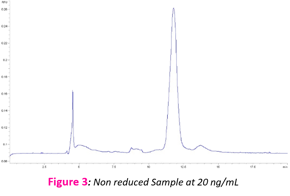
Figure 5 shows the impurity profile of a reduced and non reduced IgG standard, even apparently pure antibodies.
Experimental conditions:
Instruments | Agilent HP3D CE Picometrics ZETALIF detector Laser: 266 nm (excitation) |
Sample | IgG Standard |
LIF Capillary | Internal Diameter : 50µm Total length: 33 cm Effective length: 14 cm |
Method | •Injection: water : 1000mbars, 0.2min •Sample : 15 seconds at -15kV •Wait 0.2min in vials of water •Analysis condition: -15kV •Temperature (cassette and sample): 15°C |
Native fluorescence detection with Laser Induced Fluorescence is a simple and sensitive solution for MAbs production like impurities assay thanks to the good resolution obtained. Antibody degradation, molecular instability or manufacturing inconsistency can be easily followed with this method.
- [2] Hunt G, Nashabeh W (1999) Anal Chem.71, 2390–97,
- * LOD calculated for a ratio S/N=3
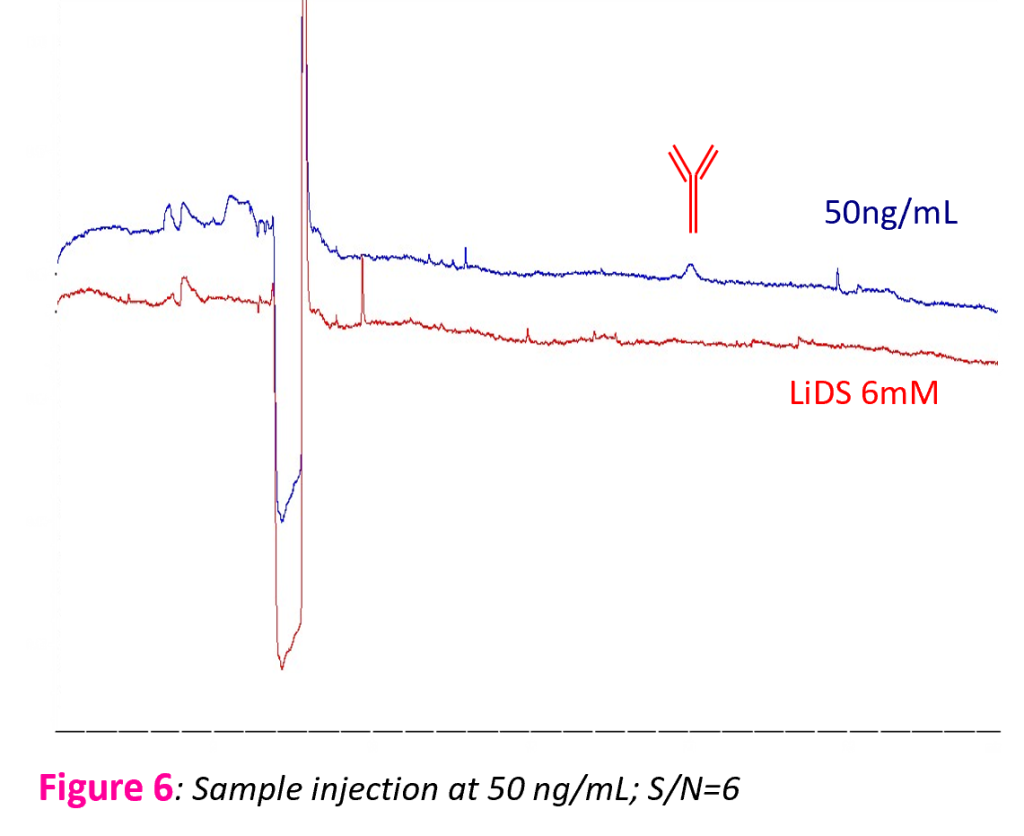
LOD* obtained with native fuorescence IgG is: 25 ng/mL
Comparison of the different detection in CE-SDS
- UV Absorbance
- Native Fluorescence
- Fluorescence derivatization
CE-SDS | UV Absorbance | Native Fluorescence | Fluorescence Derivatization | |
Wavelength nm | 210 -220 | 266 | 488 | 532 |
LOD | 500ng/ml[2] to 1µg/ml | 25ng/ml | 150pg/ml | 50pg/ml |