
µLAS technology relies on a pressure driven flow of a viscoelastic fluid in a microchannel, in which DNA is carried away and subjected to counter-electrophoresis. The combined action of the laminar, viscoelastic flow and the counter-electrophoresis produces a lift force which drives DNA molecules towards channel walls. The force intensity depends on shear stress, electric field and on the size of the DNA. When the lift force is high enough, DNA molecules are so close to the walls, where the flow velocity is low, that they crawl backward by electrophoresis. This phenomenon allows to concentrate DNA above a given size at the junction between two capillaries of sufficiently different diameters. Here, we used µLAS technology to fractionate DNA, using the applied voltage to tune the size selection threshold.
Method of Fractionnation
Instruments:
- Capillary Electrophoresis: Agilent Technologies CE7100
- Detector: Picometrics ZETALIF Laser 488nm

Key features of the method:
- Hydrodynamic injection of sample (Fig. 1A) : the stoechiometry of the sample is perfectly preserved
- No intercalating dye in the sample : best DNA quality for downstream analyses
- The hydrophilic and neutral polymer used to confer viscoelasticity is compatible with usual processes molecular biology, like PCR amplification or sequencing
CfDNA Fractionnation
Method:
A capillary of 500 µm ID was assembled end-to-end to a capillary of 50 µm ID
- Hydrodynamic injection of ~15µl of sample diluted in PVP (Fig. 1A)
- Running buffer with PVP 360 kDa 5%
- Method of concentration : 9.8mm/s and 1650V/cm (Fig. 1B)
- Method of fractionation: After concentration, the electric field is decreased at 627V/cm for 4 minutes for collecting fragments ≤ 160 bp (Fig. 1C)
- Size distribution of original and fractionated samples was determined using the BIABooster, with DNA 1K kits [1]
We used the above described method to fractionate cell-free circulating DNA, to extract DNA fragments smaller than 160 bp, which was shown to be enriched in tumoral DNA in patients with advanced cancers.
Figure 2 shows the fractionation of a typical cfDNA sample with a cut-off at 160bp using µLAS technology. The fluorescence profile of sample before fractionation is shown in red and the sample collected at same dilution in blue.
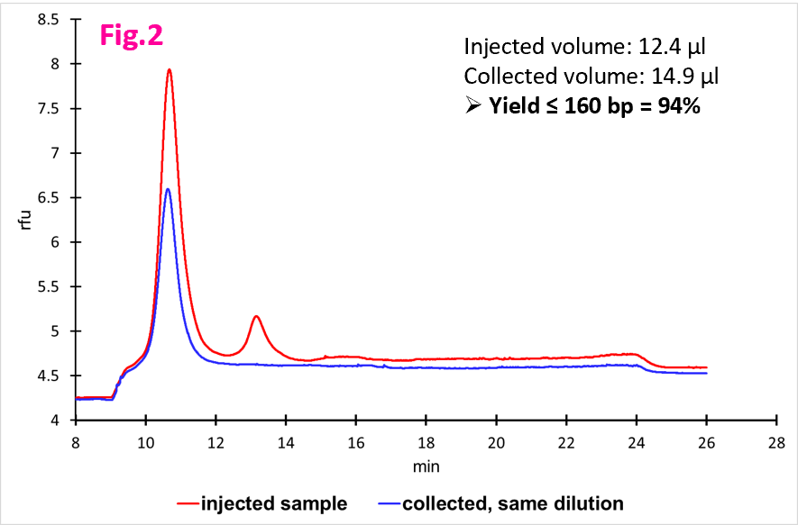

Figure 3 shows cumulated concentration (pg/µl, 10X diluted) in function of DNA size (bp): in red, the sample before fractionation and in blue, the collected sample at same dilution.
A yield of 94% of DNA ≤ 160 bp was obtained for this sample.
We have then fractionated cfDNA samples coming from pregnant women : the average yield was 70% ±10%. Original and fractionated samples were analyzed by PCR for fetal DNA; fractionation brings an enrichment factor which varied between 1.2 and 4.2.
Large DNA Fractionnation
Method:
- A capillary of 330 µm ID was assembled end-to-end to a capillary of 50 µm ID
- Hydrodynamic injection of ~1.7µl of sample
- Running buffer with PVP 10 kDa 2.5%
- Method of fractionation: 1.3mm/s, three threshold voltages of 43V/cm, 31V/cm and 24V/cm
- Size distribution of original and fractionated samples was determined using the BIABooster, with the method described in ref [2]
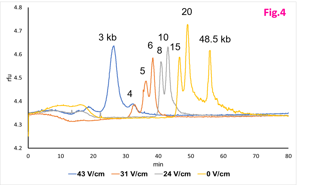
The method has also been successfully validated for the fractionation of high molecular weight DNA fragments of several kbs. Figure 4 shows the fractionation of a DNA standard into four fractions. The resulting four fractions were characterized by size selection cut-offs of 4, 7 and 12 kb. The final retained fraction plotted in yellow corresponded to the peaks of 15, 20, and 48.5 kb.
The recovered fractions can be sequenced using standard methods:
> This fractionation method has been used successfully to isolate and sequence directly a 30 kb fragment from a plant genome cut by CAS-9 assisted targeting [2].
> It has also been used to remove smaller DNA fragments of genomic DNA for long-read sequencing using the Chromium Genome Sequencing Solution from 10× Genomics (Pleasanton, CA) [3].