
µLAS technology consists in applying a combination of an electric field and pressure in a viscoelastic fluid to concentrate and separate DNA in a BIABooster capillary. It usually gives a sensitivity of 10 fg/µL [1]. In this application note, a multiple injection method is used to enhance the detection sensitivity of a weakly concentrated sample which can be injected up to ten times (10µl). A limit of detection down to 1 fg/µL for a given DNA fragment is then reached.
The method is illustrated with a circulating cell-free DNA (cfDNA) sample. cfDNA refers to extracellular DNA present in blood, coming from both normal and diseased cells. More and more studies demonstrate its potential as a noninvasive biomarker.
Method of multiple injections
Instrumentation:
- Capillary Electrophoresis: Agilent Technologies 7100 CE
- Detector: Zetalif LED 480 nm
Method:
- BIABooster with DNA 1K method [1].
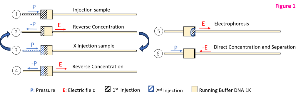
- Method consists in injecting hydrodynamically 1µl of sample and performing a reverse concentration at the inlet junction with µLAS technology (Fig 1). These steps are repeated several times up to a maximum volume injected of 10 µl. An electrophoresis is then performed to take off the whole sample from the injection chamber before concentration and separation.
cfDNA multiple injections
The method described above is used to increase the sensitivity of cfDNA purified sample.
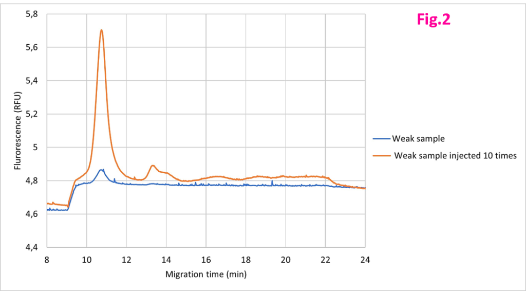
Figure 2 shows the fluorescence of DNA profile of a weakly concentrated cfDNA sample in blue and the same sample injected 10 times in orange. Sensitivity is not sufficient to have a precise detection of peaks from 200 bp to 1500 bp in the blue curve. Multiple injection method increases the accuracy of the sizing and the quantification. The size of the second and third peaks is then determined.
Figure 3 shows the multiple injections of a ladder DNA 1K (in blue, one injection; in grey, 3 injections; in pink 5 injections; in green 8 injections; in orange 10 injections.) It is possible to inject from 1 to 10 times, depending on the desired final concentration or level of accuracy.
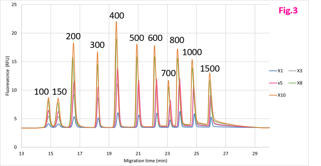
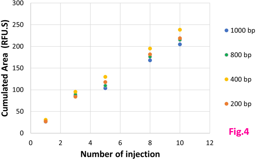
Figure 4 shows the cumulative area by size (1000 bp, 800 bp, 400 bp, 200 bp) injected one time, 3 times or 5, 8, 10 times. The increase of area is linear with a R2 close to 0.99 .
Limit of Detection
| Limit of Detection (fg/µL) |
100 bp | 13.58 |
150 bp | 5.11 |
200 bp | 3.77 |
300 bp | 3.25 |
400 bp | 1.58 |
500 bp | 1.06 |
600 bp | 0.74 |
700 bp | 0.67 |
800 bp | 0.59 |
1000 bp | 0.67 |
1500 bp | 0.82 |
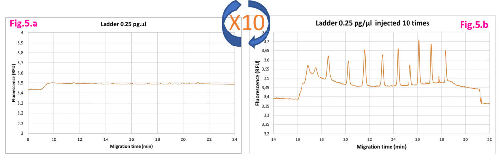
Figure 5 shows the fluorescence profile of a ladder at 0.25pg/µl injected one time and ten times.
The multiple injection method has a detection limit 10 times lower compared to the DNA 1K method.
Resistance to 10 mM Salts
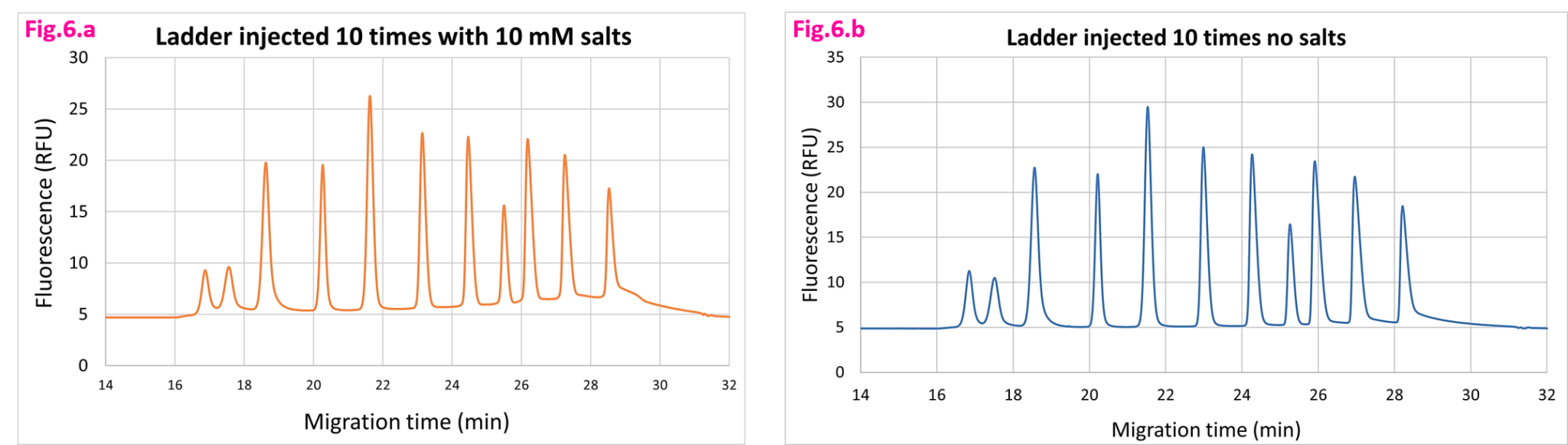
Figure 6 compares the fluorescence profile without salts and with 10mM of salts in sample. The multiple injection method supports up to 10mM of salts.
Specifications
Analytical Specifications | Value |
Sizing Range | 0.1 – 1.5 kb |
Limit of Detection (S/N = 3) | 1 fg/µl at 1kb – 10 fg/µl at 100 bp |
Sizing Accuracy | +/- 3% |
Sizing Precision | 2 % |
% RSD Area | < 20% |
Yield for 10 injections | > 70% |
Minimum Sample Volume | 15 µl |
Maximum Concentration of Salts | 10 mM |