
Pharmaceutical Antibodies are therapeutics that play an important role in controlling a broad range of diseases such as cancer, allergy, inflammation, infectious and autoimmune diseases. Monoclonal antibodies have become a fast-growing class of biopharmaceutical products.
To support analytical characterization in process development and quality control of therapeutic antibodies, capillary electrophoresis-sodium dodecyl sulfate (CE-SDS) has been recognized as an important tool in place of SDS-Page because of the ease of use and the ability to automate.
The use of 5-carboxytetramethylrhodamine succinimidyl ester (5-TAMRA.SE) and 3-(2-furoyl)-quinoline-2 carboxaldehyde (FQ) as derivatizing agents improves the sensitivity and the reproducibility of the quantification.
FQ Derivatization Method
FQ (λmax. Exc.: 480nm; λmax. Emis.: 590nm) is a fluorogenic reagent: it becomes fluorescent only upon reaction with a primary amine.
The method of derivatization is rapid and no purification after labeling is necessary.
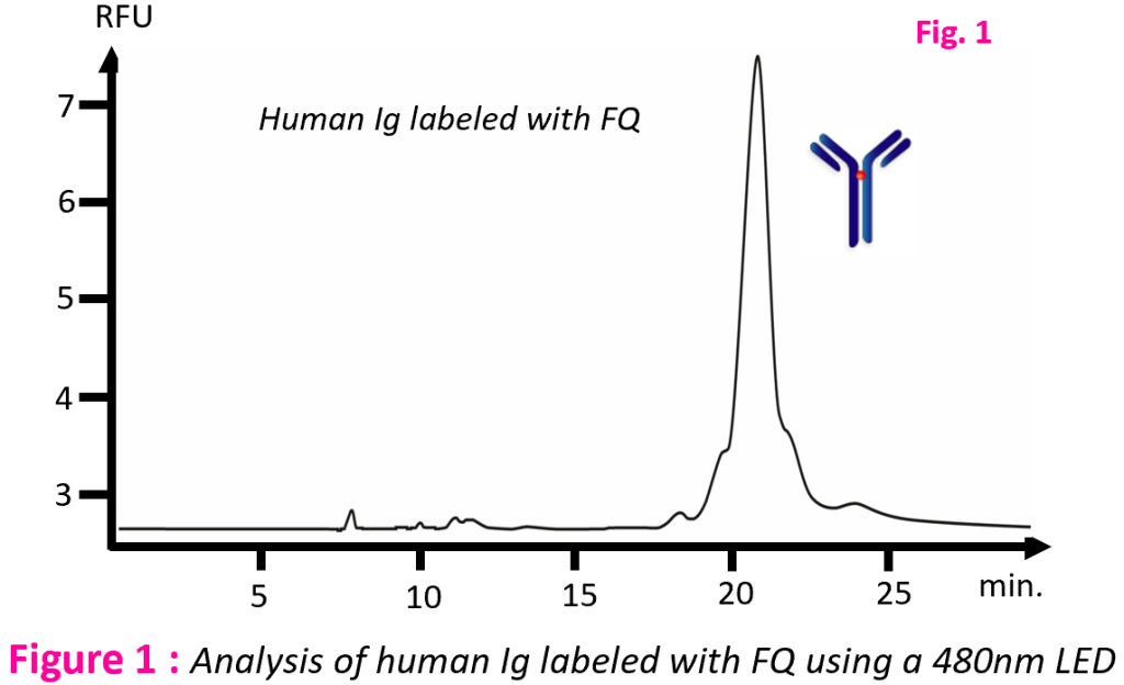
Instruments | Capillary electrophoresis:Agilent Technologies 7100 CE |
Sample | Human Ig labeled with FQ |
Labeling | rMAb (2mg/mL) was mixed in 35 µL of 0.1 M citrate-phosphate (pH 6.5), 2% SDS, 10 mM NEM, 1 mM KCN, and 25 nmol FQ; Labeling reactions were incubated for 5 min at 75℃ |
Method | – Capillary: 33 cm x 50 µm ID (effective length 19 cm) – Buffer of commercial Beckman SDS kit – Voltage: -16 kV – Injection: -10kV, 10 s – Cassette temperature : 40℃ |
To measure the limit of detection, rMAb sample was spiked with a 28kDa protein size standard at different levels as: 0.2%, 0.5%, 1%, 2%, 5%.
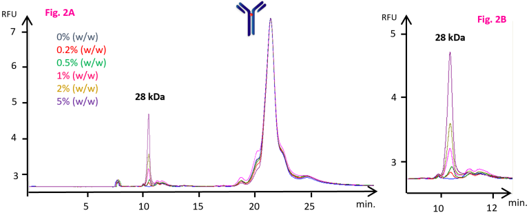
Figure 2 shows that the S/N ratio is 26 at 0.2%.
The extrapolation to a S/N ratio of 3 shows a limit of detection of 0.023% (w/w).
Figure 2A : Separation of human IgG labeled with FQ with five levels of contaminants via CE/LEDIF using a 480nm LED
Figure 2B : Extension of the egram of figure 2A between 9 and 12 minutes.
5-TAMRA.SE Derivatization Method
5-TAMRA.SE (λmax. Exc.: 550nm; λmax. Emis.: 590nm) is commonly used as a labeling reagent of mAbs for fluorescence detection as it increases the sensitivity and maintains the profile of the analyte species.
Figure 3 shows the analysis of human Ig using two LEDs (480nm and 530nm). Optimization of the excitation wavelength (530nm instead of 480nm) magnifies the S/N ratio by a factor 18.
Instruments | Capillary electrophoresis: Agilent Technologies 7100 CE Detector: Picometrics ZETALIF LED with 480nm/30nm LED and 530nm/30nm LED |
Sample | Human Ig labeled with 5-TAMRA.SE |
Labeling | rMAb samples (2mg/mL) were exchanged into 0.1 M sodium bicarbonate, pH 8.3, using a NAP-5 column. 10µL of 5-TAMRA.SE (1.4 mg/mL) dissolved in DMSO was then added to 190 µL of rmAb solution and the resultant mixture incubated for 2 h at 30℃. After incubation, 190 µL of the antibody-dye conjugate was loaded onto a second NAP-5 column and collected in 700 µL of 0.1 M sodium bicarbonate, pH 8.3. Non reduced SDS-rmAb conjugates were prepared by mixing 100 µL of the rmAb-dye conjugate and 100 µL of the CE-SDS sample buffer |
Method | Same as FQ method |
FQ derivatization | 5-TAMRA.SE derivatization | ||
Wavelength (nm) | 480 | 480 | 530 |
Derivatization method | – Fluorogenic reagent, no purification after labeling – Rapid method of labeling (5min at 75℃) | – The excess of dye should be removed with NAP-5 column – Long period of time required for labeling and purification (2 hours at 30℃ + purification) | |
Ratio S/N on the main peak | 3700 | 1400 | 25800 |
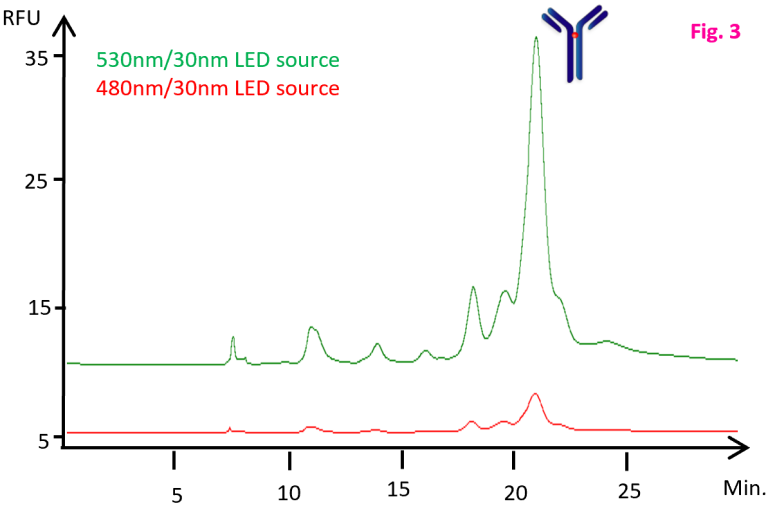
Figure 3 : Analysis of human Ig labeled with 5-TAMRA.SE using 480nm and 530nm LEDs
Conclusion:
This note describes two methods of derivatization of IgG with 5-TAMRA.SE and FQ.
The CE-LEDIF system is used to assess the purity and heterogeneity of IgG and its isoforms.
LED light source presents many advantages : less expensive, less energy consumable and more stable.